THRA Polyclonal Antibody for Western Blot, IF, ICC, IHC (P) and GS

THRA Antibody (PA1-211A) in IF
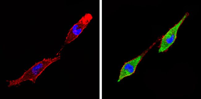
Immunofluorescent analysis of Thyroid Hormone Receptor alpha-1 (green) showing staining in the nucleus and cytoplasm of A431 cells (right) compared to a negative control without primary antibody (left). Formalin-fixed cells were permeabilized with 0.1% Triton X-100 in TBS for 5-10 minutes and blocked with 3% BSA-PBS for 30 minutes at room temperature. Cells were probed with a Thyroid Hormone Receptor alpha-1 polyclonal antibody (Product # PA1-211A) in 3% BSA-PBS at a dilution of 1:100 and incubated overnight at 4 °C in a humidified chamber. Cells were washed with PBST and incubated with a DyLight-conjugated secondary antibody in PBS at room temperature in the dark. F-actin (red) was stained with a flourescent red phalloidin and nuclei (blue) were stained with Hoechst or DAPI. Images were taken at a magnification of 60x.


