ROS 活性氧检测试剂盒

ROS 活性氧检测试剂盒

有货

ROS 活性氧检测试剂盒

品牌:Jinpan
Reactive Oxygen Species Assay Kit

MSDS

质检证书(CoA)

相似产品

货号 (SKU) 包装规格 是否现货 价格 数量
R272916-1000T 1000T 现货 ROS 活性氧检测试剂盒  

基本信息

产品名称 ROS 活性氧检测试剂盒
英文名称 Reactive Oxygen Species Assay Kit
运输条件 超低温冰袋运输

一般描述

产品组成


产品名称

包装

A液

H2DCFH-DA(10mM)

0.1mL

B液

活性氧阳性对照(Rosup,50mg/mL)

1mL


产品简介

活性氧检测试剂盒(Reactive Oxygen Species Assay Kit)是一种利用荧光探针H2DCFH-DA进行活性氧检测的试剂盒。H2DCFH-DA本身没有荧光,可以自由穿过细胞膜,进入细胞内后,可以被细胞内的酯酶水解生成DCFH。而DCFH不能通透细胞膜,从而使探针很容易被装载到细胞内。细胞内的活性氧可以氧化无荧光的DCFH生成有荧光的DCF。检测DCF的荧光就可以知道细胞内活性氧的水平。根据活细胞中荧光的产生,可以判断细胞活性氧的含量和变化。用流式细胞仪或荧光显微镜可直接观察,是一种经典的组织或活细胞中活性氧检测方法。本试剂盒提供了活性氧阳性对照试剂Rosup,以便于活性氧的检测。Rosup是一种混合物,浓度为50mg/mL。Rosup 为活性氧阳性诱导药物,根据其荧光信号强度,可分析活性氧的真正水平。

本试剂盒本底低,灵敏度高,线性范围宽,使用方便。本试剂盒可以测定1000个样品1000T(96 孔板)。

一套包含96孔板,可使用1000次。
对光敏感,注意避光保存

注意事项

1. 探针装载后,一定要洗净残余的未进入细胞内的探针,否则会导致背景较高。

2. 阳性对照Rosup 一般使用浓度为100 μM (推荐浓度100-400μM,具体依细胞类型而定)。通常刺激后0.5-4h 可观察到显著的活性氧水平升高。对于不同的细胞,活性氧阳性对照的效果可能有较大的差别。如果在刺激后30min 内观察不到活性氧的升高,可延长诱导时间或适当提高活性氧阳性对照的浓度。如果活性氧升高得过快,可缩短诱导时间或适当降低活性氧阳性对照的浓度。

3. 对于某些特别的细胞,实验过程中如果发现没有刺激的阴性对照细胞荧光也比较强, 可以按照1:2000-1:5000 稀释DCFH-DA , 使DCFH-DA 的终浓度为2-5 μM。探针装载的时间也可以根据情况在15-60 min 内适当进行调整。

4. 活性氧阳性对照(Rosup) 仅仅用于作为阳性对照的样品,并不是在每个样品中都需加入活性氧阳性对照。

5. 探针装载完毕并洗净残余探针后,可以进行激发波长的扫描和发射波长的扫描,以确认探针的装载情况是否良好。DCF的激发光谱和发射光谱请参考上图。

6. 尽量缩短探针装载后到测定所用的时间(刺激时间除外),以减少各种可能的误差。

7. 为了您的安全和健康,请穿实验服并戴一次性手套操作。

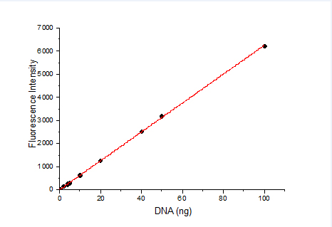
8. 定量的话要作标准曲线吧。先做一个不同浓度H2O2氧化DCFA荧光值,做一条标准曲线,X轴为H2O2浓度,y轴是荧光值,得出一个方程,在看你样品的荧光值即Y值是多少,对应的X值就是。

9. 有的细胞装载探针后细胞容易漂起来,洗细胞时实验组会吸走一部分细胞。所以种细胞时细胞量增加一倍,这样细胞紧密连接,贴壁比较牢,实验组的荧光值就高了。另外的H2DCFDA很敏感,工作液浓度要低一些,1-2μM就够啦,浓度太高容易有非特异性染色。这个探针很不稳定,一旦氧化了本底荧光值就会升高,最好工作液现用现配。

使用说明

1. 装载ROS 探针

1.1 原位装载探针(仅适用于贴壁细胞)

a) 细胞准备:检测前一天进行细胞铺板,确保检测时细胞数量小于5×10^5/ml。

b) 药物诱导:去除细胞培养液,加入无血清培养基稀释的药物处理,于37℃细胞培养箱内避光孵育,实际诱导时间由药物特性和细胞类型决定。

c) (可选)阳性对照:先用无血清培养基等稀释阳性对照(Rosup, 100 mM)到常用工作浓度100 μM,加入细胞,37℃避光孵育0.5~4 h,以提高活性氧水平,不同细胞类型存在差异。例如:HeLa 细胞需孵育30-60 min,MRC5 人胚胎成纤维细胞则需孵育90 min。

d) ROS 探针准备:探针装载前按照1:1000 用无血清培养液稀释DCFH-DA,使其终浓度为10 μM。

e) ROS 探针装载:吸除处理药物,加入适当体积稀释好的DCFH-DA 工作液。加入的体积需充分盖住细胞。例如:6孔板通常不少于1000 μL,对于96 孔板通常不少于100 μL。37℃细胞培养箱内避光孵育30 min。

f) 细胞清洗:用无血清培养液洗涤细胞1~2 次,以充分去除未进入细胞内的DCFH-DA。

1.2 收集细胞后装载探针(适用于贴壁细胞和悬浮细胞)

a) 细胞准备:按照标准方法培养细胞,必须保证检测用细胞状态。按照适当方法,清洗并收集足量的细胞。

b) 药物诱导:将收集好的细胞悬浮于适量稀释好的药物,于37℃细胞培养箱内避光孵育,实际诱导时间由药物特性和细胞类型决定。

c) (可选)阳性对照:先用无血清培养基稀释阳性对照(Rosup, 100 mM)到常用工作浓度100 μM,加入细胞,37℃避光孵育0.5~4 h 以提高活性氧水平,不同细胞类型存在差异。例如:HeLa 细胞需孵育30-60 min,MRC5 人胚胎成纤维细胞则需孵育90min。

d) ROS 探针准备:探针装载前,按照1:1000 用无血清培养液稀释DCFH-DA,使其终浓度为10 μM。

e) 探针装载:除去细胞内药物,离心收集细胞,加入稀释好的探针,使其细胞密度为1×10^6 ~ 2×10^7。

注意:细胞密度需根据后续的检测体系,检测方法,以及检测总量来进行调整。例如:对于流式分析,单管检测内细胞数目不少于10^4,也不可多于10^6。

f) 细胞清洗:用无血清细胞培养液洗涤细胞1-2 次,以充分去除未进入细胞内的DCFH-DA。

2. 荧光显微照相操作方法

a) 对贴壁生长细胞或活组织,可直接在荧光显微镜下观察;对悬浮生长细胞,取25-50 μL 细胞悬液滴到一张显微载玻片上,再盖上一张盖玻片。

b) 荧光显微镜下,选用FITC 滤光片观察荧光,去除背景观察荧光的变化。

3. 流式细胞分析操作方法

a) 对贴壁生长细胞,用胰酶消化制备成单细胞悬液;对悬浮生长细胞,直接收集细胞。用0.5-1 mL PBS 重悬细胞(0.5~1 x 10^5/ml)。

b) 选择流式细胞仪FL1 或BL1 通道,488nm 激发,测定530nm 的发射,细胞应可分成两个亚群:ROS 阴性细胞仅有很低的荧光强度,ROS 阳性细胞有较强的绿色荧光。

4.参数设置

使用488nm激发波长,525nm发射波长,实时或逐时间点检测刺激前后荧光的强弱。DCF的荧光光谱和FITC非常相似,可以用FITC的参数设置检测DCF。DCF的激发光谱和发射光谱参考下图。
ROS 活性氧检测试剂盒

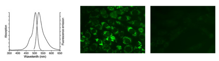
使用活性氧检测试剂盒(Reactive oxygen species assay kit)显示CHO细胞内活性氧荧光。

左图:CHO细胞用试剂盒配备的活性氧阳性对照处理;右图:正常CHO细胞。绿色荧光表明细胞活性氧急剧增加,并能显示其定位。

Product composition


Product name

Package

Liquid A

H2DCFH-DA(10mM)

0.1mL

Liquid B

Active oxygen positive control(Rosup,50mg/mL)

1mL


Product Introduction

Reactive Oxygen Species Assay Kit (Reactive Oxygen Species Assay Kit) is a kit that uses fluorescent probe H2DCFH-DA for reactive oxygen detection. H2DCFH-DA itself has no fluorescence and can freely pass through the cell membrane. After entering the cell, it can be hydrolyzed by intracellular esterase to produce DCFH. DCFH cannot penetrate the cell membrane, so that the probe is easily loaded into the cell. The reactive oxygen species in cells can oxidize non-fluorescent DCFH to produce fluorescent DCF. Detecting the fluorescence of DCF can know the level of reactive oxygen species in the cell. According to the fluorescence produced in living cells, the content and changes of reactive oxygen species can be judged. It can be directly observed with a flow cytometer or a fluorescence microscope. It is a classic method for detecting reactive oxygen species in tissues or living cells. This kit provides Rosup, a positive control reagent for active oxygen, to facilitate the detection of active oxygen. Rosup is a mixture with a concentration of 50 mg/mL. Rosup is a reactive oxygen-inducing drug. According to its fluorescence signal intensity, the true level of reactive oxygen can be analyzed.

The kit has low background, high sensitivity, wide linear range and convenient use. This kit can measure 1000 samples 1000T (96 well plate).

Precautions

1. After the probe is loaded, be sure to wash the remaining probes that have not entered the cell, otherwise it will cause a high background.

2. The positive control Rosup generally uses a concentration of 100 μM (the recommended concentration is 100-400 μM, depending on the cell type). Usually, a significant increase in reactive oxygen species can be observed 0.5-4h after stimulation. For different cells, the effect of reactive oxygen species positive control may be quite different. If no increase in active oxygen is observed within 30 minutes after stimulation, the induction time can be extended or the concentration of active oxygen positive control can be appropriately increased. If the reactive oxygen species increase too quickly, shorten the induction time or appropriately reduce the concentration of the reactive oxygen species positive control.

3. For some special cells, if the negative control cells without stimulation are found to have stronger fluorescence during the experiment, you can dilute DCFH-DA at 1:2000-1:5000 to make the final concentration of DCFH-DA 2-5 μM. The loading time of the probe can also be appropriately adjusted within 15-60 min according to the situation.

4. Active oxygen positive control (Rosup) is only used as a positive control sample, and it is not necessary to add active oxygen positive control to every sample.

5. After the probe is loaded and the remaining probes are cleaned, scan the excitation wavelength and emission wavelength to confirm whether the probe is well loaded. Please refer to the figure above for the excitation and emission spectra of DCF.

6. Try to shorten the time from probe loading to measurement (except stimulation time) to reduce all possible errors.

7. For your safety and health, please wear lab coats and disposable gloves.

8. If it is quantitative, make a standard curve. First make a different concentration of H2O2 oxidized DCFA fluorescence value, make a standard curve, the X axis is the H2O2 concentration, the y axis is the fluorescence value, and get an equation. Look at the fluorescence value of your sample, that is, the Y value, and the corresponding X value Yes.

9. After some cells are loaded with probes, the cells are easy to float up, and the experimental group will suck some of the cells when washing the cells. Therefore, the cell mass is doubled when the cells are planted, so that the cells are tightly connected and adhere to the wall more firmly, and the fluorescence value of the experimental group is higher. In addition, H2DCFDA is very sensitive. The concentration of the working solution should be lower, 1-2μM is enough. If the concentration is too high, non-specific staining may occur. This probe is very unstable. Once it is oxidized, the background fluorescence value will increase. It is best to use the working solution and prepare it.

Instructions for use

1. Load ROS probe

1.1 In-situ loading of probes (only for adherent cells)

a) Cell preparation: Plating cells the day before the test to ensure that the number of cells during the test is less than 5×10^5/ml.

b) Drug induction: Remove the cell culture medium, add the drug treatment diluted with serum-free medium, and incubate in a 37°C cell culture incubator in the dark. The actual induction time is determined by the characteristics of the drug and the cell type.

c) (Optional) Positive control: First dilute the positive control (Rosup, 100 mM) with serum-free medium, etc. to a common working concentration of 100 μM, add cells, and incubate at 37°C for 0.5 to 4 h in the dark to increase the level of reactive oxygen species , Different cell types are different. For example: HeLa cells need to be incubated for 30-60 minutes, and MRC5 human embryonic fibroblasts need to be incubated for 90 minutes.

d) ROS probe preparation: Before loading the probe, dilute DCFH-DA with serum-free culture medium at a ratio of 1:1000 to make the final concentration 10 μM.

e) ROS probe loading: aspirate the treatment drug, and add DCFH-DA working solution diluted in an appropriate volume. The added volume must cover the cells sufficiently. For example, a 6-well plate is usually no less than 1000 μL, and a 96-well plate is usually no less than 100 μL. Incubate for 30 min in a cell culture box at 37°C in the dark.

f) Cell washing: Wash the cells with serum-free culture medium 1 to 2 times to fully remove the DCFH-DA that has not entered the cells.

1.2 Load probes after collecting cells (applicable to adherent cells and suspension cells)

a) Cell preparation: Cells are cultured according to standard methods, and the cell status for testing must be guaranteed. According to the appropriate method, wash and collect enough cells.

b) Drug induction: Suspend the collected cells in an appropriate amount of diluted drug, and incubate in a 37°C cell incubator in the dark. The actual induction time is determined by the characteristics of the drug and the cell type.

c) (Optional) Positive control: First dilute the positive control (Rosup, 100 mM) with serum-free medium to a common working concentration of 100 μM, add cells, and incubate at 37°C in the dark for 0.5 to 4 hours to increase the level of reactive oxygen species. There are differences in cell types. For example: HeLa cells need to be incubated for 30-60 minutes, and MRC5 human embryonic fibroblasts need to be incubated for 90 minutes.

d) ROS probe preparation: Before loading the probe, dilute DCFH-DA with serum-free culture medium at a ratio of 1:1000 to make the final concentration 10 μM.

e) Probe loading: remove the intracellular drugs, collect the cells by centrifugation, and add the diluted probe to make the cell density 1×10^6~2×10^7.

Note: The cell density needs to be adjusted according to the subsequent detection system, detection method, and total detection volume. For example, for flow cytometry, the number of cells in a single tube is not less than 10^4 and not more than 10^6.

f) Cell washing: Wash the cells with serum-free cell culture medium 1-2 times to fully remove the DCFH-DA that has not entered the cells.

2. Fluorescence microscopy operation method

a) For adherent growth cells or living tissues, you can directly observe under a fluorescence microscope; for suspended growth cells, drop 25-50 μL of cell suspension onto a microscope slide, and then cover with a cover glass.

b) Under a fluorescence microscope, use FITC filters to observe fluorescence, and remove background to observe changes in fluorescence.

3. Flow cytometry operation method

a) For adherent growth cells, trypsinization is used to prepare a single cell suspension; for suspension growth cells, the cells are directly collected. Resuspend the cells with 0.5-1 mL PBS (0.5-1 x 10^5/ml).

b) Choose the FL1 or BL1 channel of the flow cytometer, excite at 488nm, and measure the emission at 530nm. Cells should be divided into two subgroups: ROS negative cells have only very low fluorescence intensity, and ROS positive cells have strong green fluorescence.

4. Parameter setting

Using 488nm excitation wavelength, 525nm emission wavelength, real-time or time-by-time detection of fluorescence intensity before and after stimulation. The fluorescence spectrum of DCF is very similar to FITC, and the parameters of FITC can be used to detect DCF. Refer to the figure below for the excitation and emission spectra of DCF.

ROS 活性氧检测试剂盒

Reactive oxygen species assay kit was used to display the fluorescence of reactive oxygen species in CHO cells.

Left image: CHO cells were treated with the reactive oxygen species included in the kit; right image: normal CHO cells. Green fluorescence indicates a sharp increase in cellular reactive oxygen species and can show its location.

相关属性

储存温度 避光,-20°C储存
品牌 Jinpan

PicoGreen dsDNA 定量检测试剂盒

PicoGreen dsDNA 定量检测试剂盒

有货

PicoGreen dsDNA 定量检测试剂盒

品牌:Jinpan
PicoGreen dsDNA Assay Kit

MSDS

质检证书(CoA)

相似产品

货号 (SKU) 包装规格 是否现货 价格 数量
P266245-200T×10 200T×10 期货 PicoGreen dsDNA 定量检测试剂盒  
P266245-2000T 2000T 期货 PicoGreen dsDNA 定量检测试剂盒  

基本信息

产品名称 PicoGreen dsDNA 定量检测试剂盒
英文名称 PicoGreen dsDNA Assay Kit
运输条件 冰袋运输

一般描述

产品介绍

用于 dsDNA 定量检测的 PicoGreen荧光染料

1.应用说明

在分子生物学的试验过程中,PicoGreen dsDNA 定量试剂盒是荧光检测双链 DNA 并进行定量 的一种产品,这种方法非常灵敏。常用于分子生物学技术中的:cDNA 文库的构建;用于亚克隆的  DNA 片段纯化及应用,比如进行 DNA 定量、产物扩增和引物的进一步检测。 疫苗是现代疾病预防中常用的控制方式。如今许多疫苗是细胞培养疫苗,比如重组乙肝疫苗、狂犬病疫苗等大多数疫苗都采用细胞培养的方法生产。其中,疫苗的纯化是关键问题,我们需要尽可能的去除宿主细胞DNA和宿主蛋白。假若宿主细胞的DNA和蛋白同疫苗一起注入人体将会产生不可预料的后果。

常规的DNA含量的检测方法是在260nm(A260)处测其吸光值。这种方法的主要缺点是核苷酸、单链核酸和蛋白质对信号的影响很大,并且还会受到核酸制备过程中污染物的干扰,无法区分DNA和RNA,而且这种方法不灵敏(5μg/mLdsDNA溶液A260=0.1)。PicoGreen定量检测方法简单、方便,被多家生物制品厂所选择,成为生物制品残留DNA检测的标准。

目前该方法已纳入2010年版《中国药典》

原理:

PicoGreen与DNA双链结合后才发出的荧光,无DNA不发荧光;所发荧光与DNA浓度成正比。在2010年《中国药典》中提出,PicoGreen定量DNA的方法检出限约0.3ng/ml,DNA含量在1.25-80ng/mL范围时线性较好(R2>0.99).

优点:

1)该方法可以测定来源于任何表达宿主样品中的双链DNA。

2)可以直接定量PCR扩增产物而无需从反应混合物中纯化DNA。

3)远远超出传统紫外A260的检测方法和Hoechst33258的灵敏度。

4)较高浓度的盐,尿素,乙醇,氯仿,去垢剂,蛋白或琼脂糖对测定无影响。

5)在等摩尔浓度 ssDNA 和 RNA 存在的条件下测定 dsDNA,其影响很小。

2.所需器材

• 微型荧光计;便携式荧光仪-上海互帼科学仪器有限公司HG-9型;1cm石英比色皿

• PicoGreen dsDNA 定量检测试剂盒, 1mL 单位量的试剂浓缩液足够 2mL 体积的 200 次测定。

1×TE(10mM Tris 1mM EDTA)pH8.0; 250ug/mL Sigma 小牛胸腺DNA

3.实验方案

试剂制备

PicoGreen dsDNA 定量试剂是以 1mL 的浓缩液形式保存在无水的 DMSO(二甲基亚砜)中。实 验当天,配制 2XPicoGreen 试剂的操作溶液,用 1xTE 按 1:200 的比例稀释浓缩液(10mM Tris-HCl,1mM EDTA,pH7.5)。如果要准备足够的操作溶液测定 20 个样品,可在 20mL1x TE 中加入 100μL PicoGreen dsDNA 定量试剂。由于试剂容易吸附到玻璃表面,要在塑料容器中配制。PicoGreen 试剂 见光易降解,所以应将配好的溶液用箔包住或放置暗处避光保存。

溶液最好在配制好数小时内使用,以保证最佳结果。

实验方法:

1). 标准品工作液的配制:

 Sigama小牛胸腺嘧啶DNA干粉(货号:D4522-1MG)1mg(Tris,Nacl等浓度已成标准体系),加入1mL双蒸水,配制成1mg∕mL的标准品工作液;

2). 染料工作液的配置:

6 uLPicoGreen加入1mL TE(注意:用1×TE将PicoGreen稀释200倍,现用现配,注意避光)。

3). 标准品工作液稀释:

1)母液稀释:取10ul(1mg∕mL)标准品工作液加入到990ul TE溶液中,浓度稀释成10ug∕mL,取10ul(10ug∕mL)标准品工作液加入到990ul TE溶液中,浓度稀释成100ng∕mL;

2)倍比稀释:取800ul (100ng∕mL)的标准品工作液加入到200ul TE溶液中,浓度达到80ng∕mL(药典规定:荧光染色方法DNA含量在1.25-80 ng/mL范围线性较好, 该法DNA检出限为0.3 ng/ml),取500ul(80ng∕mL)的标准品工作液加入到500ul TE溶液中,浓度稀释到40ng∕mL;依次倍比稀释,配成20ng/ml、10ng/ml、5.0ng/ml、2.5ng/ml、1.25ng/ml、0.625ng/ml的标准品溶液;

4).标准曲线的制备:倍比稀释后的各梯度标准品溶液和染料工作液各取100ul混匀,避光室温放置5min。使用FB-15型便携式荧光仪检测样品的荧光值:将混合后的溶液加入微量比色皿,确信不要在样品中引入气泡,轻轻地弹微量检测皿的外部,可以驱散气泡。以1×TE缓冲液为blank,测定样品和空白对照的荧光值;用标准品溶液的浓度(ng/ml)对应的荧光强度作直线回归,制备标准曲线。

5). 测量剩余样品的荧光值。荧光计将给出一个直接的浓度读数,数据可以用来产生DNA 浓度的标准曲线。

PicoGreen dsDNA 定量检测试剂盒


产品参数

Ex(nm) 488      Em(nm) 520

Product description

PicoGreen fluorescent dye for dsDNA quantitative detection

1. Application description

In molecular biology experiments, the PicoGreen dsDNA quantification kit is a product for fluorescent detection and quantification of double-stranded DNA. This method is very sensitive. Commonly used in molecular biology technology: cDNA library construction; DNA fragment purification and application for subcloning, such as DNA quantification, product amplification, and further primer detection. Vaccines are a commonly used control method in modern disease prevention. Many vaccines today are cell culture vaccines, such as recombinant hepatitis B vaccine, rabies vaccine and most of the vaccines are produced using cell culture methods. Among them, the purification of vaccines is a key issue, and we need to remove host cell DNA and host proteins as much as possible. If the host cell’s DNA and protein are injected into the human body together with the vaccine, it will have unpredictable consequences.

The conventional DNA content detection method is to measure its absorbance at 260nm (A260). The main disadvantage of this method is that nucleotides, single-stranded nucleic acids and proteins have a great influence on the signal, and are also interfered by contaminants in the nucleic acid preparation process, and cannot distinguish between DNA and RNA, and this method is not sensitive (5μg) /mLdsDNA solution A260=0.1). The PicoGreen quantitative detection method is simple and convenient, and has been selected by many biological product manufacturers, and has become the standard for the detection of residual DNA in biological products. At present, this method has been included in the 2010 edition of “Chinese Pharmacopoeia”

Principle:

The fluorescence emitted after PicoGreen is combined with the double strand of DNA, without DNA, does not emit fluorescence; the fluorescence emitted is proportional to the concentration of DNA. It was proposed in the Chinese Pharmacopoeia in 2010 that the detection limit of PicoGreen method for quantifying DNA is about 0.3ng/ml, and the linearity is better when the DNA content is in the range of 1.25-80ng/mL (R2>0.99).

Advantage:

1) This method can measure double-stranded DNA in samples derived from any expression host.

2) It is possible to directly quantify PCR products without purifying DNA from the reaction mixture.

3) Far beyond the traditional UV A260 detection method and the sensitivity of Hoechst33258.

4) Higher concentrations of salt, urea, ethanol, chloroform, detergent, protein or agarose have no effect on the determination.

5) Measuring dsDNA in the presence of equimolar concentrations of ssDNA and RNA has little effect.

2. Required equipment

• Mini Fluorometer; Portable Fluorometer-Shanghai Hubei Scientific Instrument Co., Ltd. HG-9; 1cm quartz cuvette

• PicoGreen dsDNA Quantitative Detection Kit, a 1mL unit volume of reagent concentrate is sufficient for 200 determinations in a volume of 2mL.

1×TE (10mM Tris 1mM EDTA) pH8.0; 250ug/mL Sigma calf thymus DNA

3. Experimental protocol

Reagent preparation

PicoGreen dsDNA quantification reagent is stored in anhydrous DMSO (dimethyl sulfoxide) in the form of a 1mL concentrated solution. On the day of the experiment, prepare the operating solution of 2XPicoGreen reagent and dilute the concentrated solution (10mM Tris-HCl, 1mM EDTA, pH7.5) with 1xTE at a ratio of 1:200. If you want to prepare enough operating solution to measure 20 samples, you can add 100μL PicoGreen dsDNA quantification reagent to 20mL1x TE. Since the reagent is easily adsorbed to the glass surface, it should be prepared in a plastic container. PicoGreen reagent is easily degraded when exposed to light, so the prepared solution should be wrapped in foil or stored in a dark place away from light.

The solution is best used within a few hours of preparation to ensure the best results.

Experimental method:

1). Preparation of standard working solution:

Sigama calf thymine DNA dry powder (article number: D4522-1MG) 1mg (Tris, Nacl and other concentrations have become standard systems), add 1mL of double distilled water to prepare 1mg/mL standard working solution;

2). Dye working solution configuration:

6 Add 1mL TE to uLPicoGreen (Note: Dilute PicoGreen 200 times with 1×TE, and use it now, please avoid light).

3). Standard working solution dilution:

(1) Mother liquor dilution: Take 10ul (1mg/mL) standard working solution and add it to 990ul TE solution, dilute to 10ug/mL, take 10ul (10ug/mL) standard working solution and add it to 990ul TE solution, concentration Dilute to 100ng∕mL;

(2) Multiple dilution: Take 800ul (100ng/mL) standard working solution and add it to 200ul TE solution, the concentration reaches 80ng/mL (pharmacopoeia stipulates: the DNA content of fluorescent staining method is linear in the range of 1.25-80 ng/mL Ok, the DNA detection limit of this method is 0.3 ng/ml). Take 500ul (80ng/mL) of standard working solution and add it to 500ul TE solution, and dilute to 40ng/mL; successively dilute by multiples and make 20ng/mL. Standard solution of ml, 10ng/ml, 5.0ng/ml, 2.5ng/ml, 1.25ng/ml, 0.625ng/ml;

4). Preparation of standard curve: take 100ul of each gradient standard solution and dye working solution diluted by multiple ratios and mix them evenly, and place them at room temperature in the dark for 5 minutes. Use the FB-15 portable fluorometer to detect the fluorescence value of the sample: add the mixed solution into the micro cuvette, make sure not to introduce air bubbles into the sample, and gently flick the outside of the micro detection cuvette to disperse the air bubbles. Use 1×TE buffer as blank to measure the fluorescence values ​​of the sample and the blank control; use the fluorescence intensity corresponding to the concentration of the standard solution (ng/ml) to make a linear regression to prepare a standard curve.

5). Measure the fluorescence value of the remaining samples. The fluorometer will give a direct concentration reading, and the data can be used to generate a standard curve of DNA concentration.
PicoGreen dsDNA 定量检测试剂盒


Product parameter

Ex(nm) 488        Em(nm) 520

相关属性

储存温度 2-8°C储存,避光,干燥
品牌 Jinpan

OligoGreen ssDNA 定量检测试剂盒

OligoGreen ssDNA 定量检测试剂盒

有货

OligoGreen ssDNA 定量检测试剂盒

品牌:Jinpan
OligoGreen ssDNA quantitative detection kit

MSDS

质检证书(CoA)

相似产品

货号 (SKU) 包装规格 是否现货 价格 数量
O273080-2000T 2000T 期货 OligoGreen ssDNA 定量检测试剂盒  
O273080-200T×10 200T×10 期货 OligoGreen ssDNA 定量检测试剂盒  

基本信息

产品名称 OligoGreen ssDNA 定量检测试剂盒
英文名称 OligoGreen ssDNA quantitative detection kit
运输条件 冰袋运输

一般描述

OliGreen寡核苷酸定量试剂是一种超灵敏的荧光核酸染料,用于定量溶液中的寡核苷酸和单链DNA(ssDNA。使用OliGreen ssDNA定量试剂从0.1到1000 ng / mL的合成24-mer(M13测序引物)线性定量。在480 nm下激发10 mm°C 10 mm比色皿中的样品。使用分光荧光计在520 nm处测量荧光发射强度,并绘制为寡核苷酸浓度的函数。寡核苷酸浓度在0到2.0 ng / mL之间获得的结果的扩大。

OliGreen Oligonucleotide Quantitative Reagent is an ultra-sensitive fluorescent nucleic acid dye that is used to quantify oligonucleotides and single-stranded DNA (ssDNA) in solution. Use OliGreen ssDNA Quantitative Reagent for synthesis from 0.1 to 1000 ng/mL 24- mer (M13 sequencing primer) linear quantification. Excite the sample in a 10 mm°C 10 mm cuvette at 480 nm. Measure the fluorescence emission intensity at 520 nm using a spectrofluorometer and plot it as a function of oligonucleotide concentration The expansion of the results obtained with oligonucleotide concentrations between 0 and 2.0 ng/mL.

相关属性

储存温度 2-8°C储存,避光,干燥
品牌 Jinpan

MTT溶液 [用于细胞增殖检测]

MTT溶液 [用于细胞增殖检测]

5.0 mg / mL in PBS

有货

MTT溶液 [用于细胞增殖检测]

品牌:Jinpan
MTT Solution [for Cell proliferation assay]

MSDS

质检证书(CoA)

相似产品

货号 (SKU) 包装规格 是否现货 价格 数量
M405849-1Set 1套 期货 MTT溶液 [用于细胞增殖检测]  

基本信息

产品名称 MTT溶液 [用于细胞增殖检测]
英文名称 MTT Solution [for Cell proliferation assay]
别名 3-(4,5-二甲基噻唑-2-基)-2,5-二苯基-2H-四唑-3-鎓溴化物溶液
英文别名 3-(4,5-Dimethylthiazol-2-yl)-2,5-diphenyl-2|H|-tetrazol-3-ium Bromide Solution
规格或纯度 5.0 mg / mL in PBS
运输条件 超低温冰袋运输

一般描述

Product description:

3-(4,5-Dimethyl-2-thiazolyl)-2,5-diphenyltetrazolium Bromide (MTT) is a type of tetrazolium salt that changes to formazan when taken up into living cells. As formazan is a non-water soluble crystal, it can be dissolved in DMSO and the absorbance at 570 nm can be used to count the number of cells. This product consists of 5.0 mg / mL MTT dissolved in PBS. When 1/10th of the medium is added to cultured cells, the live cells are properly stained.


Product Information:

Volume/Quantity : 1mL x 5

Product Form : 5.0 mg / mL in PBS

Directions for Use:

MTT solution can be added at any point in the cell culture. For best results, it should be added to cells in the logarithmic growth phase. Pre-determine the number of cells and seed 10,000 to 100,000 cells per well in a 96-well plate.

1. Seed 100 μL of cells in a 96-well plate.

2. Incubate overnight in an incubator.

3. Add 10 μL of MTT solution to each well.

4. Incubate for 2-4 hours for color reaction.

5. The medium is removed, taking care not to inhale the formazan.

6. Add 100 μL of DMSO (Product No. D0798) and dissolve the formazan.

7. Measure the absorbance at 570 nm.

Storage:
This product should be stored at -20 °C and should be protected from light. Avoid repeated freezing and thawing.

相关属性

敏感性 对光和热敏感
储存温度 避光,-20°C储存
品牌 Jinpan

刃天青 (配制的溶液) [用于细胞增殖检测]

刃天青 (配制的溶液) [用于细胞增殖检测]

有货

刃天青 (配制的溶液) [用于细胞增殖检测]

品牌:Jinpan
Resazurin (Ready-to-use solution) [for Cell proliferation assay]

MSDS

质检证书(CoA)

相似产品

货号 (SKU) 包装规格 是否现货 价格 数量
R405885-25ml 25ml 期货 刃天青 (配制的溶液) [用于细胞增殖检测]  

基本信息

产品名称 刃天青 (配制的溶液) [用于细胞增殖检测]
英文名称 Resazurin (Ready-to-use solution) [for Cell proliferation assay]
别名 刃天青 (配制的溶液) [用于细胞增殖检测]
运输条件 超低温冰袋运输

相关属性

敏感性 对热敏感
储存温度 -20°C储存
品牌 Jinpan

禽类甲型流感病毒抗体检测试剂盒 货号:274012

禽类甲型流感病毒抗体检测试剂盒 货号:274012

禽类甲型流感病毒抗体检测试剂盒

货  号:274012

产品规格:2 Test plates (strips)

原  产  地:德国

参考价格:2890 (参考价格,以实际价格为准)

优惠价格:

产品详细信息

禽类甲型流感病毒抗体检测试剂盒  2 Test plates (strips)  274012  2890元

用于检测禽类样本中甲型流感病毒的抗体

    采用ELISA方法直接对禽类甲型流感病毒进行灵敏、特异性的检测
    适用于血清、血浆样本
    检测流程采用即用型试剂,十分便利
    1.5小时内获得结果

flocktype AIV Ab采用灵敏、特异性的ELISA方法检测鸡或火鸡的血清、血浆样本中的禽类甲型流感病毒抗体。该产品能够快速、可靠的检测禽类是否感染该病毒。
flocktype AIV Ab只适用于兽医学检测。

在德国依据德国动物疫病法§17c已注册。注册号:FLI-B 435

原理
禽流感是由数种甲型流感病毒株系引起的。家禽和野生禽类都可能患禽流感。甲型流感病毒可分为低致病性或高致病性。H5和H7属于高致病性株系,可导致严重的系统性症状,即禽流感。

flocktype AIV Ab采用ELISA方法(酶联免疫吸附)和有色的即用型试剂进行检测。检测孔板包被有该病毒的重组结构蛋白。该蛋白在各禽类甲型流感病毒株系中都高度保守,免疫原性高。因此,可检测到各甲型流感血清型。

在样本孵育时,甲型流感病毒特异性抗体结合到固定化的抗原上。anti-IgY-HRP耦联物可检测结合到抗原上的抗体。加入显色底物溶液,变色反应即开始,10分钟后结束。使用分光光度计测定光密度(OD)。OD值与样本中的抗甲型流感病毒抗体的浓度相关。
操作流程
血清、血浆可在稀释后进行检测。

将存有样本的检测孔板孵育30分钟。如果含有甲型流感病毒抗体,则抗体会与孔板中的抗原结合。未结合的材料将在清洗步骤中被去除。

加入单克隆anti-IgY-HRP耦联物。未结合的耦联物被洗去。

加入TMB显色底物溶液,变色反应即开始,10分钟后结束。使用分光光度计测定光密度(OD)。获得的数据易于分析、解读。
应用
flocktype AIV Ab可检测鸡和火鸡血清、血浆样本中的甲型流感病毒的抗体。

鸡传染性支气管炎病毒抗体检测试剂盒 货号:274302

鸡传染性支气管炎病毒抗体检测试剂盒 货号:274302

鸡传染性支气管炎病毒抗体检测试剂盒

货  号:274302

产品规格:24T

原  产  地:德国

参考价格:2890 (参考价格,以实际价格为准)

优惠价格:

产品详细信息

鸡传染性支气管炎病毒抗体检测试剂盒 274302 QIAGEN

用于检测鸡样本中传染性支气管炎病毒的抗体

    采用ELISA方法,高度灵敏、特异性的检测传染性支气管炎病毒
    适用于血清、血浆样本
    检测流程采用即用型试剂,十分便利
    1.5小时内获得结果

flocktype IBV Ab采用灵敏、特异性的ELISA方法检测鸡的血清、血浆样本中的传染性支气管炎病毒的抗体。
flocktype IBV Ab只适用于兽医学检测。

在德国依据德国动物疫病法§17c已注册。注册号:FLI-B 386

原理
传染性支气管炎的临床症状与鸡的年龄、免疫接种状态、传染路径和病毒株系有关。1至4周大小的鸡会出现喘气、咳嗽、呼吸困难的症状,并伴有啰音和鼻分泌物。该疾病的致死率通常在25%至30%之间。
 
flocktype IBV Ab采用ELISA方法(酶联免疫吸附)和有色的即用型试剂进行检测。检测孔板包被有该病毒的重组核壳蛋白。该蛋白在各传染性支气管炎病毒株系中都高度保守,是主要的免疫原性肽。

在样本孵育时,传染性支气管炎病毒特异性抗体结合到固定化的抗原上。anti-IgY-HRP耦联物可检测结合到抗原上的抗体。加入显色底物溶液,变色反应即开始,10分钟后结束。使用分光光度计测定光密度(OD)。OD值与样本中的抗传染性支气管炎病毒抗体的浓度相关。

操作流程
血清、血浆可在稀释后进行检测。

样本在检测孔板中孵育30分钟。如果样本中存在传染性法氏囊病病毒的抗体,则抗体会与孔板中的抗原结合。未结合的材料将在清洗步骤中被去除。

加入anti-IgY-HRP耦联物。未结合的耦联物被洗去。

加入TMB显色底物溶液,变色反应即开始,10分钟后结束。使用分光光度计测定光密度(OD)。获得的数据易于分析、解读。
应用
flocktype IBV Ab可检测鸡血清、血浆样本中传染性支气管炎病毒的抗体。